By Nicole Golden
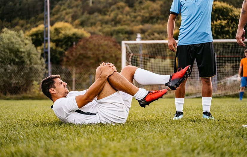
There is nothing quite like the disappointment of excitedly starting an exercise program, preparing for a new season of your favourite sport, or attempting a personal record (PR) only to find yourself injured and on the sidelines. It can also be very frustrating when that dull ache or pain becomes more bothersome as you continue your favourite activities.
Sports injuries are extremely common and will affect approximately 21 per cent of active adults (Bueno et al., 2018). In the case of youth athletes, approximately 44 per cent will suffer an injury at some point in their athletic careers (Prieto-González et al., 2021).
Sports injuries can range from mild to severe and are classified as acute or chronic (more commonly described as overuse injuries. Acute (sudden) injuries may be the first that comes to mind in sports such as soccer, football, and wrestling.
These types of injuries often prompt the athlete to seek medical attention and can sometimes be severe most commonly involving the knee (54 per cent), the hand (11 per cent), and the shoulder (7 per cent). Conversely, overuse injuries are often overlooked as they present with a gradual onset of symptoms. It is not uncommon for an athlete or active adult to not recognize they are dealing with a serious overuse injury and continue to participate in their sport without a visit to a healthcare provider (Wojtys, 2010).
What are some of the most common sports injuries? Are they preventable? How are they treated?
Sports injury prevention is chapter 13 of the NASM sports performance course (NASM-PES) and is one of the most important components of the course.
ACUTE INJURIES
TORN ACL
The ACL (anterior cruciate ligament) connects the femur to the top of the tibia. It is the most injured ligament in the knee and affects approximately 1 out of every 3500 people in the United States per year. ACL tears can be noncontact or indirect contact or contact in nature.
These can occur with a blow to the lateral portion of the knee (contact) or from poor landings when jumping, insufficient warm-ups, or a sudden uncontrolled twisting movement. Similarly, dysfunctional movement leading to poor dynamic knee stability is thought to be a modifiable risk factor. Non-contact injuries account for 60 to 70 per cent of ACL tears (Sherman et al., 2017).
ACL ruptures are more common in females from a non-contact standpoint. It is thought that the increased valgus angle of the femur contributes to more stress on the knee in female athletes or active adults. This can become an issue during landings.
Likewise, it is also possible that female athletes place higher stress on the ACL during deceleration as they are prone to favour the quadriceps during deceleration rather than the hamstrings (Evans & Nielson, 2019). Sometimes, the ACL will act to stabilize the knee when the muscles fail, however, the ACL is flimsy, and cannot hold up to high stress placed on it, leading it to tear.
SYMPTOMS/DIAGNOSIS OF TORN ACL
ACL rupture can often create a popping sound with loss of stability and swelling at the knee joint. Limited range of motion at the knee joint and difficulty walking, even if swelling is reduced, are also common signs of this injury (Sherman et al., 2017). Although this type of injury can be diagnosed with an exam from an experienced practitioner, imaging via MRI is mostly used to confirm the diagnosis (Nessler et al., 2017).
TREATMENT/REHABILITATION OF TORN ACL
Ligaments, unlike muscles, receive poor blood flow and hence heal slowly and often incompletely. Although ACL tears can be treated non-operative, most athletes or active adults who sustain this injury will opt for surgery and post-operative rehabilitation with a physical therapist. The surgery consists of grafting a piece of tendon to the remnants of the ligament to reinforce it (Sherman et al., 2017).
PREVENTING A TORN ACL
An ideal ACL tear prevention program consists of stability, plyometrics, and strength training components. The stability component should be focused on enhancing neuromuscular control, dynamic joint stability, proprioception, balance, and single-leg training. A huge portion of this type of training can be used to correct dysfunctional movement patterns that can lead to knee instability.
Plyometric training can improve landing mechanics and a component of neural control to reduce the likelihood of overstressing the ACL. Strength training is useful for strengthening the muscles supporting the joint, however, strength training alone (with no stability or plyometric component) may prove insufficient in preventing this type of injury (Nessler et al., 2017).
The NASM-OPT model provides the perfect framework for incorporating all three types of training. Including the Stabilization-Endurance and Strength-Endurance phases during some mesocycles, which both incorporate stability and plyometric components, even for healthy individuals can prove highly useful at preventing ACL tears (Clark et al., 2014).
TORN MENISCUS
The meniscus is a piece of cartilage that sits between the femur and the tibia providing a cushion to aid in shock absorption. A meniscus tear is the second most common knee injury behind ACL tears and will affect 61 out of every 100,000 people in the general population but is considerably higher in the athletic and active adult population.
Like an ACL tear, a torn meniscus can occur with a sudden trauma to the knee, twisting motion, rapid accelerating/decelerating, or high shearing forces that can occur with movements like kneeling or deep squatting under heavy loads (Raj & Bubnis, 2019). It is also important to note that as many as 40 per cent of individuals who have had a prior ACL tear will later develop a torn meniscus (Mordecai, 2014).
SYMPTOMS/DIAGNOSIS OF A TORN MENISCUS
It is not uncommon for a meniscus tear to happen at the same time as an ACL tear. The individual may describe a popping sensation with swelling. However, if swelling occurs over 24 hours after the injury, it is likely just a meniscus tear. Strangely, sometimes meniscus tears produce no symptoms or very vague symptoms such as generalized stiffness, swelling that develops over time, catching, clicking, instability in the knee, and restricted motion in the joint.
Although several tests can be done during the physical exam, imaging via MRI is the gold standard for diagnosing a meniscus tear (Raj & Bubnis, 2019).
TREATMENT/REHABILITATION OF TORN MENISCUS
Acutely, meniscus tears can be treated with R.I.C.E (rest, ice, compression, elevation) and can be treated conservatively with physical therapy and rest (long-term) or with surgery if symptoms fail to improve. Physical therapy will be targeted at improving the strength of the quadriceps (knee extensors) and range of motion.
Surgical intervention can come with an increased risk of osteoarthritis as compared to more conservative treatment. Interestingly, physical therapy can be extremely successful in the treatment of meniscus tears. Katz et al. (2013) demonstrated that in a group of patients that remained in physical therapy for 12 months, most of them regained knee function and reduction of pain like those patients who chose a surgical treatment option.
PREVENTING A TORN MENISCUS
Maintaining optimal movement patterns, thorough warm-ups, maintaining a healthy weight, good flexibility, and strength in the hip extensors, knee flexors, and knee extensors can go a long way towards the prevention of this injury.
Correction of over and underactive muscles is the first step to improving movement patterns. It is simple enough to screen for faulty movement patterns which can contribute to a meniscus tear such as poor ankle mobility and poor hip mobility. The overhead squat assessment (OHSA) can be used to screen for these movement patterns and help give the trainer a better idea of how to create a program that can prevent this type of injury (Clark et al., 2014).
Likewise, training programs should have a focus on strengthening the glutes, hamstrings, quadriceps, adductors, and abductors evenly (Zhang et al., 2017).
ANKLE SPRAIN
Sprains are simply a stretching or tear in one more ligament (the tissue that connects bone to bone). They commonly occur in the ankle, knee, wrist, or thumb, though ankle sprains are most common. In the ankle, sprains often occur from poor landings or maneuvering on an uneven surface leading to uncontrolled movements.
SYMPTOMS/DIAGNOSIS OF AN ANKLE SPRAIN
Ankle sprains can cause pain, swelling, stiffness, bruising, and limited range of motion at the ankle. They are often classified by severity as a grade I, II, III, with a grade I leading to some pain and swelling and grade III (full tear) leading to a full loss of function at the joint in addition to pain, swelling, and bruising. Diagnosis can be made clinically and with an X-ray to rule out fracture, however, the gold standard of diagnosing a sprain is an MRI as soft tissue injuries will not show up on X-rays (May Jr & Varacallo, 2020).
TREATMENT/REHABILITATION OF ANKLE SPRAIN
Rest, ice, compression, and elevation (R.I.C.E) are the first line of treatment when a sprain occurs. NSAIDS just as ibuprofen or Motrin can be used for pain. Mild sprains (i.e., grade I) will heal on their own with proper rest, but more severe sprains may require treatment.
The options for treatment are full immobilization with a cast, functional treatment which uses a bandage or brace followed by functional training to rehabilitate the joint, and surgery. The gold standard for most sprains is functional training as it helps to prevent a reoccurrence of the sprain. Manual therapy (i.e., joint mobilization and soft tissue massage) can be performed by a physical therapist followed by a program focusing on balance and strength will help the patient return to normal activity for higher grade sprains if surgery is not chosen (Martin & McGovern, 2016).
PREVENTING ANKLE SPRAINS
Neuromuscular training programs to correct ankle instability can go a long way toward the prevention of an ankle sprain or re-injury. A well-designed ankle injury prevention program will contain elements of flexibility (to restore normal range of motion), agility, balance, plyometrics, and strength training (Caldemeyer et al., 2020).
The OHSA provides a quick and easy tool to screen for ankle instability and overall ankle mobility. Pay special attention to any client with an excessive forward lean compensation. If the client has this compensation, it is recommended to elevate the client’s heels to see if the excessive forward lean is corrected. If the client’s form improves, likely there is a mobility restriction in the ankle joint (Clark et al., 2014). Ankle stability exercises can easily be included in a client’s training program via the NASM-OPT model.
STRAINS (PULLED MUSCLES)
A strain is described as tearing in the muscles or tendons that anchor those muscles to bone. Muscle strains account for roughly 10 to 55 per cent of all sports injuries. Active adults or athletes over the age of 40 are at higher risk for strains due to the ageing process in skeletal muscle tissue. Although muscle strains can occur anywhere, the most common muscles affected in athletes are muscles of the calves, hamstrings, quadriceps, or rotator cuff because they are subject to frequent acceleration and deceleration.
Strains can happen if the muscles are overstretched (especially at high speeds) overused, or severely overloaded. Muscle strains are categorized as grade I, II, or II with grade I strain representing damage to less than 5 per cent of the muscle tissue with most strength intact. Grade II strains often involve a higher proportion of muscle tissue and will cause some loss of strength. Grade III strains often involve rupture at the tendon and full loss of strength which may make surgery necessary (Maffulli et al., 2014).
SYMPTOMS/DIAGNOSIS OF PULLED MUSCLES
Muscle strains can cause extreme soreness, bruising, stiffness, or a feeling of extreme tightness, and in higher-grade strains, loss of strength and sometimes indentation under the skin. Muscle strains can be acute (sudden) or chronic. They may start as a nagging soreness and get worse over time, or a high-grade strain could happen suddenly. Oftentimes a clinical exam and/or ultrasound can be enough to diagnose and grade a strain though MRI may be necessary in some cases (Maffulli et al., 2014).
TREATMENT/REHABILITATION
Grade I strains will often heal on their own with gentle stretching and rest. Physical therapy is recommended for grade II and sometimes grade III muscle strains, however, if the strain occurs at the tendon, surgery may be needed to reattach the muscle to the bone to regain full function. The R.I.C.E protocol is recommended along with avoiding lengthening the muscle in the first 3 to 7 days after injury.
The second stage of treatment involves isometric training and active flexibility training assuming there is no significant pain. The third stage of rehabilitation will involve strength training. The fourth stage will include sports-specific exercise (if the injured is an athlete) including plyometric training (Fernandes et al., 2011).
PREVENTING PULLED MUSCLES
Proper warmup, optimal movement patterns (including muscle flexibility), good core stability, and good control over movements are the best ways to prevent muscle strain. A certified personal trainer must check their client for muscle imbalances before designing a training program to address these imbalances before they can lead to significant injuries (McCall et al., 2020).
Likewise, a thorough warm-up including low-intensity physical activity and dynamic stretching is recommended before playing a sport or completing an intense workout. This will help increase blood flow to the muscles, make them more pliable, and mimic the movements that will occur later in the activity (Clark et al., 2014). These steps can help to reduce the risk of muscle strain.
BURSITIS
A bursa is a fluid-filled sac located next to the tendons in some of the major joints which helps to reduce the friction of the joints moving. There is over 150 bursae in the human body. Bursitis is the term that describes inflammation of the bursa sac and can occur in any of the bursa sacs, but most commonly in the hip, knee, shoulder, ankle, or elbow.
Bursitis can occur because of a trauma to the bursa sac, repetitive motion of the joint, prolonged pressure on the joint (i.e., someone who spends a lot of time on their knees or resting on their elbows), through systemic inflammation, or infection. Typically, bursitis is a temporary condition that will not lead to permanent damage unless it is left untreated (Williams & Sternard, 2019).
SYMPTOMS/DIAGNOSIS OF BURSITIS
Oftentimes, bursitis can be diagnosed clinically by an experienced practitioner, but imaging such as ultrasound or MRI may be used especially if another type of injury needs to be ruled out. Bursitis may be acute or chronic and will often present with pain and stiffness in the joint with or without visible swelling. In some cases, the skin near the joint will feel hot to the touch (Williams & Sternard, 2019).
TREATMENT/REHABILITATION OF BURSITIS
Bursitis will often heal on its own without intervention, however, if bursitis occurred because of overuse, limiting the inciting activity for some time may be recommended. Physical therapy may be warranted and if so, therapy will focus on maintaining range of motion in the affected joint along with exercises to strengthen the muscles crossing that joint. Antibiotics will be prescribed if the infection is the cause and fluid may be removed from the bursa sac if swelling is excessive (National Center for Biotechnology, 2018).
PREVENTING BURSITIS
Athletes can be at risk for bursitis. For example, swimmers and baseball players may be at risk for shoulder bursitis, hockey players, runners, cyclists for hip bursitis, etc. Training programs that focus on joint stability can be very helpful in preventing bursitis as well as proper periodization, paying close attention to clients/athletes at risk for overuse injuries (National Center for Biotechnology, 2018).
THE OPT MODEL AND TREATING SPORTS INJURIES
Sports injuries occur because the involved body part was asked to do something it was not ready or able to do. Poor conditioning, inadequate training, insufficient warm-ups, poor flexibility, poor neuromuscular control, faulty movement patterns, and deficiencies in strength are risk factors for all these potential injuries. Dynamic movement assessments and proper use of the OPT model/periodization can be powerful weapons in the fight against sports injuries.
The OHSA can catch many faulty movement patterns and allow the fitness professional to apply the OPT model and correct any muscle imbalances contributing to the faulty movement pattern. A periodized program with elements of flexibility, balance, stability, endurance, strength, and power will provide a well-rounded training protocol to properly condition athletes or active adults for their favourite sports or activities.
The OPT model also provides an excellent framework from which a trainer can have conversations with their clients about the importance of all these training elements, proper warmups, and optimal movement patterns to educate them to make better decisions about preparation for activity.
REFERENCES
Bueno, A. M., Pilgaard, M., Hulme, A., Forsberg, P., Ramskov, D., Damsted, C., & Nielsen, R. O. (2018). Injury prevalence across sports: a descriptive analysis on a representative sample of the Danish population. Injury Epidemiology, 5. https://doi.org/10.1186/s40621-018-0136-0
Caldemeyer, L. E., Brown, S. M., & Mulcahey, M. K. (2020). Neuromuscular training for the prevention of ankle sprains in female athletes: a systematic review. The Physician and Sportsmedicine, 1–7. https://doi.org/10.1080/00913847.2020.1732246
Evans, J., & Nielson, J. l. (2019, March 8). Anterior Cruciate Ligament (ACL) Knee Injuries. Nih.gov; StatPearls Publishing. https://www.ncbi.nlm.nih.gov/books/NBK499848/

