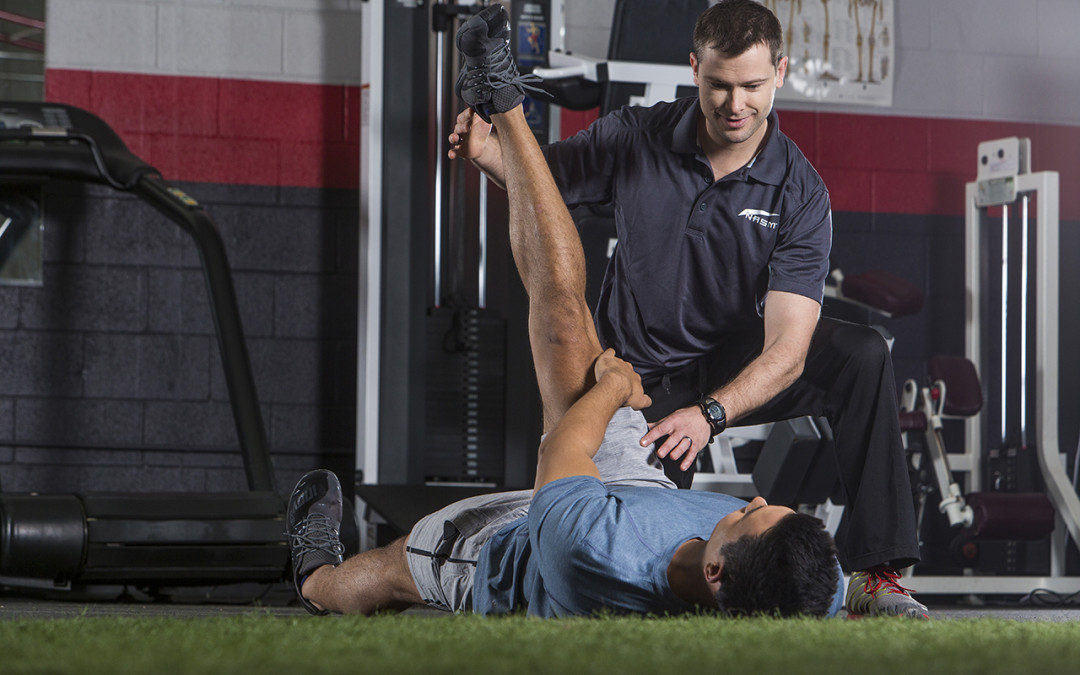
Corrective exercise uses a systematic process that involves identifying neuromusculoskeletal dysfunction, developing a plan of action and integrating a corrective strategy. This process requires knowledge and application of an integrated assessment process in order to determine the appropriate program design and exercise techniques. Here we’ll look at the why and how we assess.
Why do we assess?
- Establish a baseline/starting point
- Create realistic expectations
- Discover the specific GOALS and NEEDS of each client
- Create individualized exercise programs that are systematic and progressive
- Create value in the services we offer
- Establish ourselves as knowledgeable
- Help ensure client accountability
- Consistency = CREDIBILITY
If you are not assessing, you’re guessing.
Using the SOAP acronym can be helpful for analyzing clients and determining the appropriate program design. SOAP stands for:
- Subjective
- Objective
- Assessment
- Plan
Subjective information can be gathered using a pre-participation screening tool such as a general health history and a health-risk appraisal such as the PAR-Q (Physical Activity Readiness Questionnaire). These tools can help to identify pertinent information such as:
- Occupation
- Lifestyle
- Medical history
- Past injuries
- Surgeries
- Medications
- Dietary habits
- Exercise history
As with any exercise program design, one should start with a Needs Analysis in order to gain an understanding of what is required for the activity or sport. This should include:
- What is the basic energy system involved?
- What are the movements that must be trained?
- What are the most common injury sites?
- A biomechanical assessment
(Kraemer, 1984)
Objective information typically involves data that we can quantify and use to evaluate progress. This can include:
- Weight/Height
- Vital signs (blood pressure and pulse)
- Body composition
- Circumference measurements
- Static posture analysis
- Movement screen
- Range of motion
- Muscle testing
- Upper body strength endurance (e.g., push-up test)
- Lower body strength endurance (e.g., wall squat test)
- Sub Max VO2 (e.g., 3 minute step test)
Your Assessment will be based on the data collected from the Subjective and Objective information, which will ultimately be used to design a Plan (program design).
Kinetic Chain Assessments
A kinetic chain assessment is designed to identify dysfunction within the human movement system (HMS):
- Altered length-tension relationships of soft tissues (muscles, ligaments, tendons and fascia)
- Altered force-couple relationships (compensatory movement)
- Altered arthrokinematics (joint dysfunction)
Dysfunction in the HMS will lead to:
- Altered sensorimotor integration
- Altered neuromuscular efficiency
- Tissue fatigue and breakdown (cumulative injury cycle)
A streamlined assessment of the Kinetic Chain should include:
- Static postural assessment
- Dynamic movement screen (e.g., overhead squat assessment)
- Range of motion testing*
- Manual muscle testing*
Static Postural Assessment
Janda, a Czech neurologist, identified predictable patterns of muscle imbalance where some muscles become shortened/overactive and others become lengthened/underactive. He labeled these as: Upper Crossed Syndrome, Lower Crossed Syndrome, and Pronation Distortion Syndrome. These can be identified through a static postural assessment, by viewing the client from the anterior, lateral and posterior positions and systematically at each of the five kinetic chain checkpoints:
- Feet and ankles
- Knees
- Lumbo-pelvic-hip (LPHC) complex
- Shoulders
- Head/cervical spine
Upper Crossed Syndrome
- Characterized by: Rounded shoulders and a forward head posture. This pattern is common in individuals who sit a lot or who develop pattern overload from uni-dimensional exercise
- Short Muscles: Pectoralis major and minor, latissimus dorsi, teres major, upper trapezius, levator scapulae, sternocleidomastoid, scalenes
- Lengthened Muscles: Lower and middle trapezius, serratus anterior, rhomboids, teres minor, infraspinatus, posterior deltoid, and deep cervical flexors
- Common injuries: Rotator cuff impingement, shoulder instability, biceps tendonitis, thoracic outlet syndrome, headaches
Lower Crossed Syndrome
- Characterized by: Increased lumbar lordosis and an anterior pelvic tilt
- Short Muscles: Iliopsoas, rectus femoris, tensor fascia latae, piriformis, adductors, hamstrings, erector spinae, gastocnemius, soleus
- Lengthened Muscles: Gluteus maximus, gluteus medius, VMO, transversus abdominus, multifidus, internal oblique, anterior and posterior tibialis
- Common injuries: Hamstring strains, anterior knee pain, low back pain
Pronation Distortion Syndrome
- Characterized by: Excessive foot pronation, genu valgus and poor ankle flexibility
- Short Muscles: Peroneals, gastrocnemius, soleus, iliotibial band, hamstrings, adductors, iliopsoas
- Lengthened Muscles: Posterior tibialis, flexor digitorum longus, flexor hallicus longus, anterior, tibialis, posterior tibialis, vastus medialis, gluteus medius, gluteus maximus
- Common Injury Patterns: Plantar fasciitis, posterior tibialis tendonitis (shin splints), anterior, knee pain, low back pain
(Page, 2010)
Dynamic Movement Screen
The Overhead Squat Assessment is designed to assess dynamic flexibility, core strength, balance and overall neuromuscular efficiency. As with the static postural assessment, this should be a systematic process observed from the anterior, lateral and posterior positions, noting compensations at each of the five major Kinetic Chain Checkpoints. These compensations can signify over and under active muscles, abnormal force-couple relationships and joint dysfunction.
Overhead Squat Assessment Protocol
- Barefoot
- Feet shoulder width apart and pointed straight ahead in a neutral position
- Raise arms overhead, with elbows fully extended
- Squat to chair height and then return to start position
- 5 repetitions in anterior, lateral and posterior positions
NASM CES Solutions Table (Please refer to NASM CES Overhead Squat Solutions Table)
As you can see from the Solutions Table, there are a number of compensations characterized by potentially over and underactive muscles. By integrating range of motion and manual muscle testing, the precise muscles and joints can be isolated, streamlining the process and helping to make the program design more accurate and effective.
Range of Motion Testing
Range of motion assessment looks at the amount of motion available at a specific joint. Active range of motion occurs through voluntary contraction by the client and can be observed through the overhead squat. Passive range of motion is performed without the assistance the client and provides information about joint play and end feel.
Range of motion testing in a clinical setting often involves using a device such as a goniometer or inclinometer in order to quantify joint motion by measuring degrees.
As a personal trainer, an alternative would be to evaluate motion at the major joints as follows:
- Functional Non-Painful (FN)- Normal pain free motion
- Functional Painful (FP)- Normal motion that is painful
- Dysfunctional Painful (DP)- Abnormal motion that is painful
- Dysfunctional Non-painful (DN)- Abnormal motion that is not painful
If a movement causes pain, refer to the appropriate specialist. As a trainer, you should be looking for Dysfunctional Non-Painful (DN) movements.
The NASM Essentials of Corrective Exercise Training is a useful resource for the normal range of values for each muscle. Doing a visual comparison between sides is also helpful.
Regional Interdependence
Regional interdependence is the concept that seemingly unrelated impairments in a remote anatomical region may contribute to, or be associated with an area of pain. For example, clients who complain of low back pain or discomfort may actually be suffering from dysfunction at the ankle, hip or knee joints. By focusing corrective exercise strategies at the most Dysfunctional Non-Painful movement impairments (using the NASM CEx model- Inhibit, Lengthen, Activate, Integrate), many common problems affecting the foot and ankle, low back, knees, shoulders and neck can be addressed in a fitness setting.
(Wainner, 2007)
Remember, when in doubt, refer out!
Manual Muscle Testing
Muscle testing is an art and a science. There are a number of factors that can cause a muscle to test weak. Essentially, muscles must be properly activated by the nervous system in order to produce internal tension to overcome an external force.
NASM has developed a 3-point grading system and manual muscle testing process:
| Numerical Score | Level of Strength |
| 3 | Normal |
| 2 | Compensates (recruits other muscles) |
| 1 | Weak |
NASM 2-Step Manual Muscle Testing Process:
| Step 1 | Step 2 |
| ● Place muscle in shortened position or to point of joint compensation.● Ask client to hold that position while applying pressure.● Grade the client’s strength (3,2,1)● If client can hold position without compensation, then muscle is strong.● If muscle is weak or compensates, move to step 2 | ● Place muscle in mid range and retest strength.● If muscle is normal in mid range, there may be opposing muscle overactivity or joint hypomobility- inhibit and lengthen those opposing muscles.● If the muscle is weak or compensates in mid-range position, the muscle is likely weak. * |
*There can be a number of reasons for a weak muscle. As a trainer, you can try reactivation and reintegration techniques. If these fail to work, refer out.
Key Take-Home Points
Optimum program design and a streamlined assessment involves:
- Subjective information (e.g., PAR-Q, Health History)
- A Needs Analysis
- Objective data
- Static posture
- Dynamic Movement Screen
- Range of Motion Testing
- Manual Muscle Testing
- An Assessment (e.g., per the NASM- CES Solutions Table)
- Exercise selection based on the above per the NASM- CEx Model:
- Inhibit
- Lengthen
- Activate
- Integrate
* Disclaimer: Check with your state laws regarding the scope of practice for fitness trainers to perform passive range of motion and manual muscle testing techniques on clients.
References
Clark,M.A., & Lucett, S.C. (Eds.). (2010). NASM Essentials of Corrective Exercise Training. Baltimore, MD: Lippincott Williams & Wilkins.
Clark, M.A., Sutton, B.G., Lucett, S.C. (2014). NASM Essentials of Personal Fitness Training. 4th Edition, Revised. Burlington, MA: Jones and Bartlett Learning.
Kraemer, W.J. (1984). Exercise prescription: Needs analysis. Strength & Conditioning Journal, 6(5), 47-47.
Page, P., Frank, C., & Lardner, R. (2010). Assessment and Treatment of Muscle Imbalance: The Janda Approach. Champaign, IL: Human Kinetics.
Wainner, R. S., Whitman, J. M., Cleland, J. A., & Flynn, T. W. (2007). Regional interdependence: a musculoskeletal examination model whose time has come Journal of Orthopaedic & Sports Physical Therapy, 37(11), 658-660.

